Clinical picture
Pyruvate kinase deficiency (PKD) of red blood cells is a rare, inherited and severe form of chronic anaemia, and is a type of haemolytic anaemia. These disorders are characterised by a rapid breakdown (lysis) of blood (haemoglobin). In PKD, the malfunctioning of the enzyme pyruvate kinase (PK) is the cause of that blood breakdown.
There are several metabolic diseases in which the metabolism of pyruvate is disrupted. Pyruvate kinase ensures that the red blood cells are supplied with sufficient energy. Due to the lack of this enzyme, the red blood cells become damaged and obtain an abnormal shape. As a result of this change, the cells are more quickly removed from the blood by the liver and spleen. The bone marrow cannot keep up with the request for new blood cells and this causes anaemia.
In the Netherlands, 60 people are currently diagnosed with PKD. For many other countries, the number is unknown.
Symptoms
The characteristics of pyruvate kinase deficiency differ for each patient. Some patients experience only some mild complaints, others show very serious symptoms and need to be treated in hospital. The most common symptoms presented by patients with PKD are:
- Fatigue, weakness and paleness due to a shortage of red blood cells.
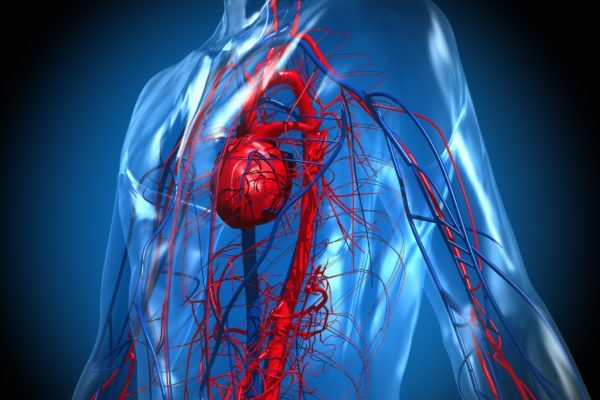
- Irregular heartbeat because the heart has to work harder to pump the reduced amount of oxygen in the blood.
- Chronic pain in bones and muscles, as a result of the accelerated blood production or blood breakdown, the iron overload or even of the drugs against iron overload.
- Jaundice: when red blood cells are demolished, haemoglobin is released into the bloodstream. The haemoglobin is degraded in the gallbladder into a yellow waste product called bilirubin. This allows skin and eyes to get a yellowish colour and also leads to a darker colour of the urine.
- Pain in the upper abdomen: due to the increased level of bilirubin in the blood that is drained via the bile duct, painful gallstones can develop.
- Low resistance to infectious diseases: particularly young PKD patients are more susceptible than their healthy peers and also have more difficulty recovering.
- Enlarged spleen.
However, there are other complications that may occur:
- Iron stacking: the accelerated degradation of red blood cells and many blood transfusions may lead to an iron surplus (iron overload). This mainly causes problems in the liver and heart, but it can also disrupt hormone production and affect the joints. It is treated with medication (chelation therapy).
- Pancreatitis due to the large flow of bilirubin: it results in pain in the middle upper abdomen and is treated with medication.
- Gilbert’s syndrome: a condition in which the bile ducts have difficulties in removing bilirubin. In combination with PKD it leads to extreme yellow vision, severe fatigue and pain attacks of the bile ducts.
Cause
Pyruvate kinase deficiency is a congenital blood disorder and inherited in an autosomal recessive manner. Autosomal means that it is not sex-linked, which means that men and women have an equal chance of inheriting it from their parents. If both parents are carriers, they have a 25% (1 in 4) chance of passing on the deficiency to their child, a 25% chance that the child has no abnormal PK gene and a 50% chance that the child will be a carrier. People who are carriers can only pass on the disease if their partner is also a carrier of the deficiency.
In most cases, both parents have no symptoms. They are called ‘healthy carriers’ of one different PK gene and have one normal gene. This normal gene ensures the production of sufficient pyruvate kinase. Only when both genes are abnormal, does this condition arise, with the enzyme being insufficiently produced, or not produced entirely. Due to the lack of this enzyme, the red blood cells become damaged and take on an abnormal shape.
Normally red blood cells circulate in the bloodstream for approximately 120 days; the abnormal cells only survive for a few days. The bone marrow cannot produce sufficient new red blood cells quickly enough to replace the damaged cells. This creates a shortage of red blood cells, or anaemia.
Diagnosis
The diagnosis of PKD is primarily based on the PK level in the blood, as well as the haemoglobin (Hb) content, both of which will be lower in patients with PKD, compared to a healthy patient. In PKD patients, this is between 4 and 7 mmol/L. In addition, genetic tests can also be carried out. Over 200 different mutations have been identified, but it is not yet clear which mutations have an effect on the severity of the disease.
The diagnosis is based on the following studies:
- Structured patient history, taking into account the signs and symptoms the patient reports.
- Physical examination.
- Blood tests:
- Complete blood count, Hb and bilirubin content, PK activity.
- Reticulocytes (young red blood cells) count to determine the production rate of new red blood cells in the bone marrow. This has been increased in haemolytic anaemia due to the accelerated degradation of the blood cells.
- Blood smears to investigate the shape of red blood cells.
- Liver function test: to determine the level of haptoglobin and bilirubin. When the red blood cells break down, the iron is bound from the cells to the haptoglobin protein. A low haptoglobin level is an indication for an increased breakdown of red blood cells.
- Genetic test for missing or mutated genes in the DNA that are associated in PKD.
Treatment
The treatment of pyruvate kinase deficiency consists of preventing and reducing the symptoms associated with chronic anaemia. Available treatments are:
- Exchange transfusion: especially when the bilirubin level is high in new-born babies, because this might damage the brain. During transfusion a part of the patients’ blood is drained by transfusion and at the same time the same amount of healthy (donor) blood is administered. This lowers the bilirubin content.
- Blood transfusion (at a low Hb level (anaemia))
- Chelation therapy (deferrization): many blood transfusions may lead to iron overload.
- Vitamins (including vitamin D and calcium), by means of healthy food.
- Folic acid to support the production of red blood cells.
- Spleen removal (splenectomy) may help to keep the Hb level more stable, requiring fewer transfusions. Due to the increased risk of infections in young children, this procedure is not performed in children younger than 2 years.
- Antibiotics and vaccinations due to the increased risk of infections in case of a spleen removal. The spleen plays an important role in fighting infections. Patients must be vaccinated with the pneumococcal vaccine against pneumonia, followed by chronic antibiotic treatment (penicillin) to prevent infections.
- Removal of gallbladder and gallstones: a large amount of bilirubin that is released during the degradation of red blood cells is sometimes not properly processed by the liver and gallbladder. The patient will turn yellow (jaundice) and may develop gallstones that causes a lot of pain.
Finally, there are some therapies (under development) that can cure patients. These therapies are still under consideration and not yet part of the treatment guidelines:
- Stem cell transplantation.
- Gene therapy in which the mutation in the PK gene of the stem cells of the bone marrow is undone and the enzyme does work.
Additional information
Patient organisations
Related articles
Links
Clinical picture

Symptoms

Cause

Diagnosis

Treatment
