Clinical picture
Langerhans cell histiocytosis (LCH) is characterised by the proliferation of Langerhans cells in the skin, bones and bone marrow. This proliferation interferes with the functioning of the tissues and organs. Langerhans cells are immune cells that have an important role in the immune system. When the Langerhans cells mature sufficiently, they are released into the blood, after which they spread through the body.
In patients with LCH, the Langerhans cells no longer enter the bloodstream. They remain at the location where they mature and this causes an accumulation of Langerhans cells. These cells start to attack normal functioning cells and cause local inflammation, called a granuloma. In this granuloma, so-called giant cells also arise because the Langerhans cells merge.
There are different types of LCH:
- Local LCH: the accumulation of Langerhans cells is limited to a specific location in the body, usually the bone. Minimal treatment is required.
- Generalised LCH: accumulation occurs at multiple locations in the body including the bones, bone marrow, lymph nodes, liver, spleen, lungs and / or brain. This type is life-threatening and usually affects very young children.
- Fading LCH: a variant of LCH that eventually disappears. During the active stage of the disease, damage is done to various tissues/organs around the body.
Langerhans cell histiocytosis is also called histiocytosis X, eosinophil granuloma, Hand-Schüller-Christian disease or Letterer-Siwe disease.
How many patients have LCH?
Langerhans cell histiocytosis predominately occurs in children up to 15 years of age, especially in children between the age of 5 and 10. It is estimated that 1 in 200,000 children are diagnosed with LCH each year.
Because LCH is very rare in adults, the incidence is difficult to estimate. Research estimates that 1 in 560,000 adults are diagnosed each year. The condition is twice as often present in men as in women. Particularly in younger children, the prognosis of LCH is very poor.
Symptoms
Because the location of Langerhans cell histiocytosis may differ for each patient, the symptoms also vary. Most of the patients’ experience fever, sickness and weight loss. Other symptoms can be:
- Formation of lumps in the bones accompanied by local pain complaints. Especially seen in the skull, but also in the jaw, pelvis, thigh, ribs and vertebrae. These lumps may result in bone abnormalities and a decreased bone density.
- Chronic discharge from the ear. This can result in hearing loss and balance problems.
- Redness and swelling of the eye: LCH in the bones of the eye socket may lead to double and poor vision.
- Eczema-like skin abnormalities
- Diabetes insipidus: this condition is present in 1 out of 4 children and is accompanied by a lot of thirst and a lots of urination.
- A disturbed production pattern of hormones:
- A deficiency of thyroid hormone causes energy loss, slowness and problems with the bowel movement.
- A growth hormone deficiency causes reduced growth and an increase in abdominal fat and excess weight.
- A shortage of sex hormones causes the absence of puberty and especially in girls, the lack of menstruation.
- Reduced adrenal stimulating hormone (ACTH) levels causes energy loss and susceptibility to infection.
- An increase in the hormone prolactin results in milk production and milk flow from the breasts in girls.
- Sleeping problems, especially in patients with LCH near the brain.
- Change in body temperature.
- Coordination disorders, mood disorders, behavioural problems and epileptic seizures.
- Anaemia: resulting in paleness, fatigue, dizziness and shortness of breath.
- Deficiency of platelets: more sensitive to bruises, longer haemorrhages and spontaneous nose bleeding.
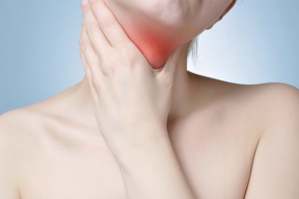
- Enlarged lymph nodes.
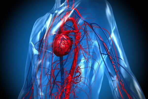
- Jaundice and diarrhoea due to malfunctioning liver and spleen. May be accompanied by dark urine, loss of appetite and itching all over the body.
- Lung abnormalities such as shortness of breath, persistent cough and pain in the ribs. Patients are also more susceptible to recurrent pneumonitis.
Cause
Although the exact cause of LCH is still unknown, research suggests that the BRAF-gene plays a role in the development of the disease. A mutation in this gene can only be found in abnormal Langerhans cells and not in other cells. The BRAF gene normally ensures that cells do not start to divide uncontrollably. Due to an error in the gene, uncontrolled growth and division in possible. An inflammatory reaction, such as a viral infection, can be the trigger of this disruption.
Diagnosis
Langerhans cell histiocytosis is a relatively rare condition. Therefore, it may take a while before the diagnosis is confirmed. The diagnosis can often be made by:
- Structured patient history, taking into the account the symptoms of the patient.
- Physical examination.
- Blood tests.
- Examination of the affected tissue.
- Bone marrow examination for abnormal Langerhans cells.
- Defining the skeleton status (more sensitive in comparison to bone scan), possibly supplemented with a CT scan or MRI study on lesions in the bones.
- Ultrasound of the lower abdomen.
- Detection of the faulty BRAFV600E gene.
Treatment
LCH treatment during childhood depends on the affected organ and location. After diagnosis, a distinction is made between low and high risk Langerhans cell histiocytosis. Patients with low-risk LCH only have LCH in the bones or skin. If the LCH is present in the liver, spleen or bone marrow, it is considered as high risk LCH. This last group needs a more extensive treatment than the first group to be able to heal.
Treatments can include:
- Local steroids cream for skin-LCH.
- Chemotherapy (vinblastine) combined with an anti-inflammatory prednisone at localisations around and on the skull.
- Operation, especially with local LCH in the bone.
- Treatment with prednisone during surgery of the affected bone.
- In generalised LCH, other medications are often used in addition to vinblastine and prednisone, such as 6-mercaptopurine and methotrexate.
- If the complaints do not reduce after a few weeks of treatment, heavier types of chemotherapy, such as cladribine and cytarabine, can be used.
- Stem cell transplantation.
- Radiation therapy to inhibit disease progression is applied when LCH is present in the vertebrae because it presses on the spinal cord and may cause paraplegia.
- Hormone replacement therapy:
- Desmopressin in diabetes insipidus.
- Levothyroxine in case of a deficiency of thyroid hormone.
- Injections with growth hormone.
- Contraceptive pill or testosterone tablets in case of a shortage of sex hormones.
- Cortisol tablets in case of a deficiency in adrenal stimulating hormone.
- Targeted therapy with kinase and BRAF inhibitors, as well as monoclonal antibodies can be used as a major reagent in patients with recurrent disease for whom no other options are available.