Cincinnati Children’s – HLH | What is Hemophagocytic Lymphohistiocytosis? (English)
(Familial) haemophagocytic lymphohistiocytosis (HLH or FHLH) is a rare and potentially life-threatening disease that occurs primarily in very young children, but can also affect adults.
Histiocytes and lymphocytes are cells of the immune system that help to fight infections. Both cells can be classified as white blood cells. Patients with HLH have histiocytes and lymphocytes with a defect and therefore are not able to respond effectively to the external threat.
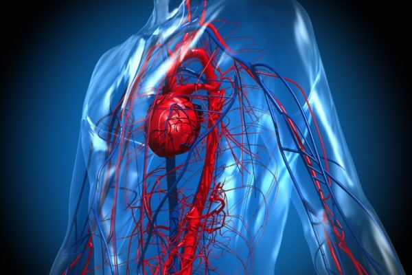
Because of the malfunctioning of these cells, more cells are attracted to the site of infection and therefore causes an accumulation of activated T-lymphocytes and histiocytes. These cells secrete cytokines (substances involved in the immune system) in a large amount, resulting in the activation and recruitment of even more white blood cells. The cells penetrate into healthy tissue and multiply, causing damage to all kinds of organs. This causes inflammation with possible symptoms: swelling, redness, heat, pain and loss of function. Depending on the location of the inflammation, damage occurs to all kinds of tissues and organs, such as bone marrow, lymph nodes, liver, spleen, skin, meninges, spinal cord and, in rare cases, the brain tissue.
The main symptoms of haemophagocytic lymphohistiocytosis are persistent fever and an enlarged liver and spleen (hepatosplenomegaly). Patients may also experience respiratory or gastrointestinal problems. Other symptoms may include:
There are two types of HLH: an inherited (genetic) type, also called primary HLH, and a reactive type called secondary HLH. Hereditary HLH can be subdivided into familial HLH (FHLH) and HLH as a result of an immune deficiency disorder.
Primary HLH is an autosomal recessive disease. This means that 2 defective genes are necessary to develop the disease: one paternal and one maternal gene. If both parents are carriers of an affected HLH gene, they can pass on the disease but do not have the disease itself. If a child inherits one healthy, functional gene and one gene with a mutation, the child will be a carrier of the disease, similar to their parents. However, in 25% of cases, a child is born that has inherited both defective genes from the parents, causing the disease to develop.
In secondary HLH, the disease occurs in response to another underlying condition, usually an infection. The most common infection that causes secondary HLH is the Epstein Barr virus (EBV). However, other causes, like infections, autoimmune diseases and malignant diseases, can lead to the development of HLH.
Diagnosing the familial type of HLH is possible by clinical research. If a very young child is diagnosed with HLH, it immediately points in the direction of the primary variant (FHLH). When patients are diagnosed after the age of 6, in most of the cases this means that they do not have FHLH. The medical history of the family is also used in the diagnostic process. If another child in the family also has the disease, there is an indication for the familial variant. However, if the disease is caused by an infection, there is still a chance that the patient has the familial type of the disease.
The diagnosis HLH can be made by:
Genetic research into genetic background of the disease is also possible. Five subtypes of FHLH are distinguished on the basis of the type of mutation. The genes affected by HLH are involved in the cell-destroying function of immune cells.
Hereditary HLH can be fatal within 2 months after diagnosis when untreated. Therefore, rapid treatment is desirable. The goal of treatment at HLH is to reduce the symptoms (remission) and get the disease under control. But even if this succeeds, the results in the familial form of the disease are only temporary. When the treatment is discontinued, the symptoms will return.
The treatment of patients with primary HLH can consist of:
In the case of secondary HLH, the treatment depends on the cause. When the course of the disease is mild, symptomatic treatments are sufficient. If possible, the underlying cause of the disease is identified and treated. If the condition cannot be controlled, a treatment like in the case of primary HLH is required.