Patiëntenvereniging Erdheim Chester Disease Global Alliance
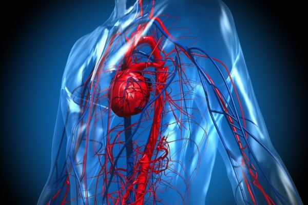
Erdheim-Chester disease (ECD), also termed a non-Langerhans-cell histiocytosis, is a very rare disease characterised by the abnormal multiplication of a specific type of white blood cell called a histiocyte (or tissue macrophages). The disease involves an infiltration of lipid-laden macrophages, multinucleated giant cells, an inflammatory infiltrate of lymphocytes and histiocytes in the bone marrow, and a generalized sclerosis of the long bones. The excess production of histiocytes (histiocytosis) leads to inflammation that can damage organs and tissues throughout the body, causing them to become thickened, dense, and scarred (fibrotic). This tissue damage may lead to organ failure.
The signs and symptoms of Erdheim-Chester disease usually appear between the ages of 40 and 70, although the disorder can occur at any age. It affects both men (60% of reported cases) and women (40%). The severity of the condition varies widely. Some affected individuals have few or no associated health problems, while others have severe complications that can be life-threatening.
Erdheim-Chester disease can involve many different systems in the body and most often affects the long bones. Therefore bone pain is the most frequent symptom associated with ECD and mainly affects the lower limbs, knees and ankles. More than 50% of cases have kidney, skin, brain and lung involvement. Symptoms include:
The following symptoms may indicate disease involvement but are not necessarily diagnostic. The disease varies greatly from patient to patient and some, but not all of these symptoms may be present.
• Bone pain, sometimes associated with muscle pain. This may be specific to one area or more widespread.
• Lower back pain, reduced kidney function/painful or difficult urination.
• Abdominal or lower back pain, painful or difficult urination due to affected retroperitoneum (a part of the internal abdominal wall).
• Skin or eyelids: rash or soft, fatty, yellow bumps.
• Brain: difficulty with coordination, staggering gait, slurred speech, behavior disorders, and rapid, involuntary eye movement (nystagmus).
• Dry cough, shortness of breath with exercise.
• Eye pain and redness, bulging eyes, difficulty with vision including double vision.
• Excessive thirst and urination (Diabetes Insipidus).
• Shortness of breath, fatigue, and swelling of feet, ankles, and lower legs.
• Weight loss, fever, muscle and joint aches, night sweats, weakness/fatigue, and increased tendency to get infections.
The specific cause of Erdheim-Chester Disease is unknown. However, more than half of people with Erdheim-Chester disease have a specific mutation in the BRAF gene. Mutations in other genes are also thought to be involved in this disorder.
The BRAF gene provides instructions for making a protein that helps transmit chemical signals from outside the cell to the cell’s nucleus. These signals regulate the growth and division (proliferation) of cells, the process by which cells mature to carry out specific functions (differentiation), cell movement (migration), and the self-destruction of cells (apoptosis).
The BRAF gene mutation that causes ECD is somatic, which means that it occurs during a person’s lifetime and is present only in certain cells. The mutation occurs in histiocytes or in immature precursor cells that will develop into histiocytes. This mutation leads to production of a BRAF protein that is abnormally active, therefore disrupting regulation of cell growth and division. The unregulated overproduction of histiocytes results in their accumulation in the body’s tissues and organs.
Because it is so rare, Erdheim-Chester disease is often difficult to diagnose. Patients may go for months and even years after symptoms start until they are properly diagnosed. This disease is sometimes mistaken for Langerhans cell histiocytosis (LCH). A diagnosis of ECD is made based upon a thorough:
• Physical examination, history of symptoms, past illnesses, etc.
• Blood tests: To evaluate function of the internal organs such as heart and kidneys.
• Biopsy: involves taking a small piece of the affected tissue and looking for foamy histiocytosis with signs of inflammation and something called Touton-type giant cells.
• Neurological examination: Testing of ability to walk, muscle testing, coordination, etc.
• X-ray: This can be performed of the lungs or the bones to determine if there are abnormalities.
• Bone scan: This is a type of x-ray that looks for bone lesions. A small amount of radioactive material is injected into a vein, collects in any abnormal parts of the bones, and is shown on a scanner.
• CT scan: This is an x-ray that takes a number of detailed pictures at different angles inside the body to create a continuous picture. Dye may be injected into a vein or swallowed to help find any abnormalities.
• MRI: This imaging study uses magnets to excite atoms within cells of the body, which then emit radio waves, and are subsequently detected by the scanner. This detection allows for the visualisation of these targeted cells. A substance called gadolinium may be injected into a vein.
• PET scan: A small amount of radioactive sugar is injected into a vein, and diseased cells show up brighter on the scanner.
• Ultrasound: High-energy sound waves are bounced off organs and tissue and make echoes, which form a picture of the internal body.
• Electrocardiogram: This is a tracing of the heart rate and rhythm.
• Echocardiogram: This uses sounds waves to provide pictures of the heart in order to evaluate blood flow and heart function.
• Genetic testing: the BRAF V600E gene mutation has been found in about half of patients with ECD.
Because Chester-Erdheim is a very rare disease, no large studies have been performed. Therefore, no treatment plan has been established that is widely accepted. However, various treatments have been used with limited success. These treatments are intended to control the symptoms and growth of the disease. Treatments include:
• Surgical debulking to decrease symptoms caused by masses of histiocytes.
• High-dose corticosteroid therapy (prednisone) to reduce inflammation in the body.
• Ciclosporin.
• Immunotherapy to restore the ability of the immune system such as Interferon-α.
• Chemotherapy to control the over-production of histiocytes This may include vinblastine, vincristine, cyclophosphamide, doxorubicin, and cladribine (2CdA). Other drugs such as methotrexate, imatinib, tamoxifen, azathioprine, and mycophenolate mofetil have also been used.
• BRAF inhibitors.
• Radiation therapy using high-energy rays to destroy rapidly increasing histiocytes.
Uncovering Rare Disease: Erdheim-Chester Disease (ECD Global Alliance