The Gaston Baele Memorial Lecture, named after the first President of the Board of the Belgian Society on Thrombosis and Haemostasis (BSTH), is held annually during the BSTH meeting. For the edition of 2022, Professor Kristin Jochmans from the University Hospital Brussels was elected to present this lecture. Prof. Jochmans focussed on antithrombin (AT), our key natural coagulant, and AT deficiency. The presentation walked us through the diverse molecular functions of AT, touched upon the diagnosis and clinical presentations of AT deficiency, including a discussion of the causes for its underdiagnosis.
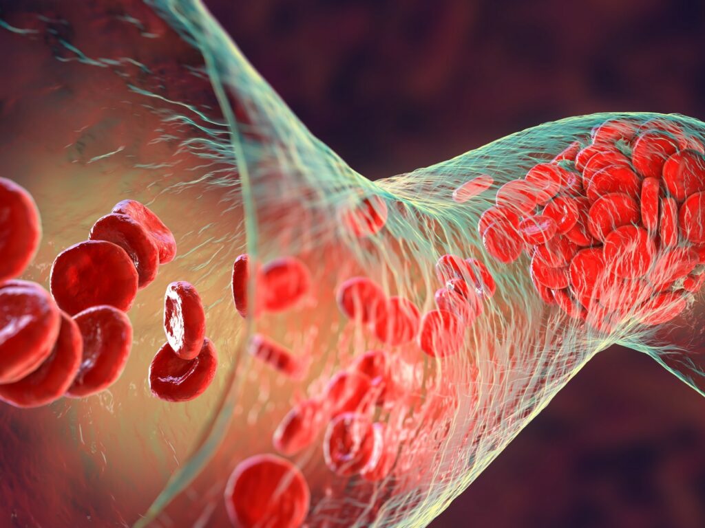
Antithrombin (AT) is a single-chain plasma glycoprotein encoded by the gene SERPINC1 that acts as a key natural anticoagulant. AT mainly inhibits coagulation factors IIa (thrombin) and Xa, but also interacts with factors IXa and XIIa, trypsin, plasmin and kallikrein. Remarkably, its activity is increased by >1000-fold in the presence of heparin. AT circulates in two isoforms in the plasma; isoforms a and b. a-AT represents 90-95% of the AT in plasma and is N-glycosylated at Asn128, 167, 187 and 224. b-AT, which makes up 5-10% of the AT in the blood is a similar molecule except that is not glycosylated at position Asn167, and has a higher affinity for heparin. The best known function of AT is anticoagulation. In this AT binds to different protease (e.g. thrombin), rendering them inactive. Heparan sulfate proteoglycans (HSPGs) are molecules that are similar to heparin and can be found on the vessel wall. These molecules are able to activate AT to avoid thrombotic events at this level. In addition to this, AT (mainly the b-AT form) is also involved in anti-inflammatory signalling. In fact, the binding of AT to specific 3-OS vascular HSPGs induces prostacyclin production by endothelial cells and other anti-inflammatory signals.1 Finally, AT also seems to exert an anti-bacterial and anti-viral effect.1–3
Clinical presentation and diagnosis
Inherited AT deficiency is a severe form of thrombophilia, characterised by an overall increased risk for a first venous thromboembolism (VTE) (about 14-fold compared to the general population). These thromboembolisms commonly occur in the deep and superficial leg veins, or in the lungs (pulmonary embolism). Nevertheless, thrombotic events may also occur in mesenteric, renal, or cerebral veins or affect arteries. AT can be diagnosed by functional assays that analyse anti-Xa or anti-IIa activity, progressive activity or immunological assays. In all these tests, an AT activity below 80% is considered to be deficient. Crossed immune-electrophoresis can be performed to analyse the subtype: type I deficiency (lower concentration of the antigen) or type II (normal concentration, but the molecule is dysfunctional). Type II deficiencies can be further divided depending on the localisation of the genetic alteration: heparin binding site (HBS), reactive site (RS) or a combination of both (pleiotropic effect, PE).4 Mutations are found in up to 80% of the cases of patients with inherited AT deficiency and mainly consist of point mutations and small INDELS.
In terms of obstetrical complications, AT is associated with an increased risk for pregnancy-related VTE. In a study including 189 pregnant women with type I AT deficiency, 10.5% suffered a VTE. Although there was a small numerical difference between the groups who received heparin and those who did not (7.0% vs. 11.6%), this difference was not significant, meaning that VTE developed irrespective of whether the patient received prophylactic heparin or not.5 In a Hungarian study with 64 pregnant women, 33% of women with type I deficiency developed a VTE. This study also analysed the effect on VTE of the AT Budapest 3 mutation (p.Leu131Phe), a founder type II HBS mutation in the Balkans, which can have a homozygous or heterozygous presentation. In total, 15% of the patients developed a VTE in the homozygous type II HBS group, and none in the heterozygous one. Birth rate was 100% for patients with type I deficiency, 94% for those with heterozygous type II HBS Budapest 3, and only 8% in patients with homozygous type II HBS Budapest 3.6 Another study observed a birth rate of 32% in patients with type II HBS Budapest, a higher value but still poor compared to the 100% in type I.7
Limitations of the current assays & underdiagnosis
AT deficiency appears to be underdiagnosed, partly because routine functional methods have many variables (incubation time, heparin concentration, type of serum, etc.) that can influence the results. The need to create more sensitive methods is, however, still under debate, since over-sensitive methods may generate off-target effects. Additionally, an AT deficiency can be transient. In fact, Bravo-Pérez et al. showed that some AT mutations are only present at certain timepoints, possibly related to external factors (e.g. alcohol intake or fever). This study analysed 444 consecutive cases with an AT activity of <80% in at least one sample. Patients were categorised as having a consecutive (CD, AT<80% in all samples) or a transient deficiency (TD, AT≥80% in at least one sample).8 In TD patients, 48 SERPINC1 variants were found (27/48 were AT Cambridge II [p.Ala416Ser] or AT Dublin [VAI30Glu], together with some hypoglycosylation problems.8 In another study, eight of 30 patients with AT deficiency but without SERPINC1 defects had AT forms with reduced molecular weight and negative charge, which suggests abnormal N-glycosylation. Of these, 5/8 had an intermittent AT deficiency with hypoglycosylation, all associated with moderate alcohol intake. Genetic analyses, including while exome sequencing, revealed recurrent mutations in phosphomannomutase 2 (PMM2) and other genes involved in the N-glycosylation pathway.9 Another study revealed two new SERPINC1 variants (p.Asn224His and p.Glu227Lys) in four patients with early and recurrent thrombosis and normal AT activity at first screening. In these patients, the a glycoform was normal, while the b isoform was abolished, which resulted in an impaired or absent N-glycosylation of Asn224. Remarkably, heating the sample for one hour at 41°C resulted in a decrease in the activity of anti-FXa. Taken together, these results suggest that alterations in N-glycosylation result in transient loss of function depending on external factors like increase in body temperature (fever) or intake of alcohol.10
AT is a versatile molecule with many roles, including anti-coagulation and anti-inflammatory signalling (mainly bythe b form). Inherited AT deficiency is characterised by an overall increased VTE risk. This risk seems to be markedly increased during pregnancy, especially in patients with homozygous type II HBS p.Leu131Phe-Budapest 3 AT. Additionally, external factors, like fever or alcohol, can induce a transient AT deficiency in patients, typically with alterations in N-glycosylation. The discovery of the different functions for a and b isoforms may mark the beginning of new potential therapeutic strategies in patients with AT, exploiting the beneficial effects of AT and identifying patient groups with possible benefits from AT supplementation.
References
